
2026 - 1 Issue

Case report

Punctate Inner Pachychoroidopathy Associated with Choroidal Neovascularization. A Case Report

Full Text links

Summary

Aims: To report a case of punctate inner choroidopathy (PIC) with pachychoroid disease features and active choroidal neovascular membrane.

Materials and methods: Case report

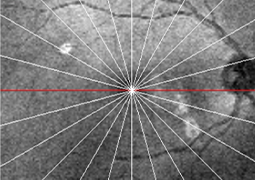
Results: A 33-year-old female patient with a history of myopic neovascular membrane in the right eye (OD), who had received multiple doses of intravitreal Aflibercept, consulted our retina service. Best-corrected visual acuity was 20/40 in OD. Fundus examination revealed small, well-defined, yellow-gray spots with subretinal fluid limited to the posterior pole. Optical coherence tomography scans from the OD showed subretinal fluid and a hyperreflective material between the epithelium/Bruch membrane (RPE/ BrM) complex, associated with loss of normal choroidal architecture and focal conformational choroidal excavation. There was also diffuse choroidal thickening in the macula, with pachyvessels compressing the inner choroid. Fundus autofluorescence showed active PIC lesions. With these findings, the diagnosis of punctate inner pachychoroidopathy associated with active choroidal neovascular membrane was made and Aflibercept therapy was restarted.

Conclusions: A subset of patients with PIC exhibits features associated with pachychoroid disease. This subtype of PIC is determined by unique demographics, multimodal image findings, and complications that differ from classic PIC, due to the potential influence of choroidal venous insufficiency on PIC manifestations and secondary complications.

References

  1. Ahnood D, Madhusudhan S, Tsaloumas MD, Waheed NK, Keane PA, Denniston AK. Punctate inner choroidopathy: A review. Surv Ophthalmol. 2017 Mar-Apr;62(2):113-126. Epub 2016 Oct 15. doi: 10.1016/j.survophthal.2016.10.003
  2. Standardization of Uveitis Nomenclature (SUN) Working Group. Classification Criteria for Punctate Inner Choroiditis. Am J Ophthalmol. 2021 Aug;228:275-280. Epub 2021 Apr 15. doi: 10.1016/j.ajo.2021.03.046
  3. Li M, Zhang X, Wen F. The Fundus Autofluorescence Spectrum of Punctate Inner Choroidopathy. J Ophthalmol. 2015;2015:202097. Epub 2015 Jul 22. doi: 10.1155/2015/202097
  4. Vance SK, Khan S, Klancnik JM, Freund KB. Characteristic spectral-domain optical coherence tomography findings of multifocal choroiditis. Retina. 2011 Apr;31(4):717-723. Epub 2015 Jul 22. doi: 10.1097/IAE.0b013e318203c1ef
  5. Ruiz-Medrano J, Montero JA, Flores-Moreno I, Arias L, García-Layana A, Ruiz-Moreno JM. Myopic maculopathy: Current status and proposal for a new classification and grading system (ATN). Prog Retin Eye Res. 2019 Mar;69:80-115. Epub 2018 Nov 1. doi: 10.1016/j.preteyeres
  6. Hady SK, Xie S, Freund KB, et al. Prevalence and characteristics of multifocal choroiditis/punctate inner choroidopathy in pathologic myopia eyes with patchy atrophy. Retina. 2022 Apr 1;42(4):669-678. doi: 10.1097/IAE.0000000000003383
  7. Cheung CMG, Lee WK, Koizumi H, Dansingani K, Lai TYY, Freund KB. Pachychoroid disease. Eye (Lond). 2019 Jan;33(1):14-33. Epub 2018 Jul 11. doi: 10.1038/s41433-018-0158-4
  8. Ramtohul P, Freund KB, Parodi MB, et al. Punctate inner pachychoroidopathy: Demographic and Clinical Features of Inner Choroidal Inflammation in Eyes with Pachychoroid Disease. Retina. 2023 Nov 1;43(11):1960-1970. doi: 10.1097/IAE.0000000000003926
  9. Kim H, Woo SJ, Kim YK, et al. Focal choroidal excavation in multifocal choroiditis and punctate inner choroidopathy. Ophthalmology. 2015;122:1534-1535.
  10. Ghadiali Q, Dansingani KK, Freund KB. focal choroidal excavation and choroidal neovascularization with associated pachychoroid. Retin Cases Brief Rep. 2016 Autumn;10(4):293-296.