
2025 - 3 Issue

Original article

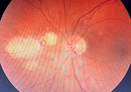
T-Cells Response in Experimental Autoimmune Uveitis of Varying Severity

Full Text links

Summary

Aim: to investigate the dynamics of the T-cell immune response in rabbits with experimental autoimmune uveitis (EAU) of varying severity.

Materials and Methods: The experiment involved two groups of Chinchilla rabbits (15 rabbits in each group). The model of EAU was created. The clinical picture of intraocular inflammation of varying severity was assessed. The determination of the level of white blood cells (WBC), lymphocytes (Lymphs), CD3+, CD4+, CD8+, and CD16+ in the blood of rabbits was conducted.

Results: Group I – moderate and severe uveitis, Group II – uveitis of mild severity. WBC, Lymphs, CD3+, CD4+, CD16+ were elevated and statistically significant in both groups of animals compared to control parameters on all days of the experiment (3, 7, 10, 14, 21 days) (p < 0.001). CD8+ level had a significantly lower count than the control one (p < 0.001). When comparing the two groups, the immune response was more active in Group I, and the number of immune cells did not return to normal by the end of the experiment.

Conclusion: In the case of EAU, the immune response is characterized by the activation of the T-cell immune system, with the intensity of this response depending on the severity of the clinical presentation of uveitis. Various degrees of clinical severity in EAU were obtained using an experimental model employed in our study. A rapid response of the immune system helps to establish a diagnosis and predict the severity of autoimmune uveitis.

References

  1. Foster CS, Vitale AT. Diagnosis and treatment of uveitis. 2nd ed. New Delhi: Jaypee Brothers Medical Publishers; 2013. p.1276.
  2. Khramenko NI, Konovalova NV, Usov VYa, Velychko LM., Bogdanova OV. Immunity status and expression of molecular markers (ICAM‑1, CD5, CD25, CD95) on lymphocytes of patients with recurrent anterior uveitis complicated by macular edema. Graefes Arch Clin Exp Ophthalmol. 2022;261:423-431.
  3. Khramenko NI, Velychko LM, Bogdanova OV, Konovalova NV, Iu. O. Zhuravok IuO. Sensitivity of peripheral blood lymphocytes to adrenaline and acetylcholine in patients with primary and recurrent posterior uveitis. J.ophthalmol (Ukraine). 2022;4:3-11.
  4. Jabs DA, Nussenblatt RB, Rosenbaum JT. Standardization of uveitis nomenclature for reporting clinical data. Results of the First International Workshop. Am J Ophthalmol. 2005;140:509-516.
  5. Caspi RR, Cohen IR, Miller A. Experimental autoimmune uveoretinitis – rat and mouse. Autoimmune Disease Models: A Guidebook. Academic Press. 1994. p.57-81.
  6. Peyman GA, Paque JT, Meisels HI, Bennett TO. Postoperative endophthalmitis: A comparison of methods for treatment and prophylaxis with gentamicin. Ophthalmic Surg. 1975;6(1):26-35.
  7. Dick AD, Cheng YF, McKinnon A, Liversidge J, Forrester JV. Nasal administration of retinal antigens suppresses the inflammatory response in experimental allergic uveoretinitis. A preliminary report of intranasal induction of tolerance with retinal antigens. Br J Ophthalmol. 1993;77:171-175.
  8. Kempen JH, Altaweel MM, Holbrook JT, Jabs DA, Sugar EA. The multicenter uveitis steroid treatment trial: rationale, design, and baseline characteristics. Am J Ophthalmol. 2010;149(4):550-561.
  9. Jabs DA, Dick A, Doucette JT, et al. Interobserver agreement among uveitis experts on uveitic diagnoses: the Standard of Uveitis Nomenclature Experience. Am J Ophthalmol. 2018;186:19-24.
  10. McNeil R. Grading of ocular inflammation in uveitis: an overview. Eye News. 2016;22(5):1-4.
  11. Thurau SR, Chan C-C, Nussenblatt RB, Caspi RR. Oral tolerance in a murine model of relapsing experimental autoimmuneuveoretinitis (EAU): induction of protective tolerance in primed animals. Clin Exp Immunol. 1997;109:370-376.
  12. Kanda Y. Investigation of the freely available easy-to-use software ‘EZR’ for medical statistics. Bone Marrow Transplant. 2013;48:452-458.
  13. Huryanov VG, Lyakh YuYe, Pariy VD, et al. Handbook of biostatistics. analysis of medical research results using EZR (R-statistics) [in Ukrainian]. Kyiv: Vistka; 2018. p.208.
  14. Streilein JW. Ocular immune privilege: the eye takes a dim but practical view of immunity and inflammation. J LeukocBiol. 2003;74:179-185.
  15. Merida S, Palacios E, Navea A, Bosch-Morell F. Macrophages and uveitis in experimental animal models. Med Inflam. 2015;2015:1-10.
  16. Caspi RR. A look at autoimmunity and inflammation in the eye. J Clin Invest. 2010;120(9):3073-83.
  17. Sonoda KH, Stein-Streilein J. Ocular immune privilege and CD1d-reactive natural killer T cells. Cornea. 2002;21:33-8.
  18. Ohta K, Wiggert B, Yamagami S, Taylor AW, Streilein JW. Analysis of immunomodulatory activities of aqueous humor from eyes of mice with experimental autoimmune uveitis. J Immunol. 2000;164:1185-92.
  19. Taylor AW. Ocular immune privilege. Eye. 2009;23:1885-9.
  20. Masli S, Vega JL. Ocular immune privilege sites. Methods Mol Biol 2011;677:449-58.
  21. Agarwal RK, Rachel R Caspi RR. Rodent models of experimental autoimmune uveitis. Methods Mol Med. 2004;102:395-419.
  22. Tang J, Zhu W, Silver PB, Su SB, Chan CC, Caspi RR. Autoimmune uveitis elicited with antigen-pulsed dendritic cells has a distinct clinical signature and is driven by unique effector mechanisms: initial encounter with autoantigen defines disease phenotype. J Immunol. 2007;178(9):5578-87.
  23. de Smet MD, Chan CC. Regulation of ocular inflammation - what experimental and human studies have taught us. Prog Retin Eye Res. 2011;20(6):761-97.
  24. Chan СС, Caspi RR, Ni M, Leake WC, Wiggert B, Chader GJ, et al. Pathology of experimental autoimmune uveoretinitis in mice. J Autoimmun. 1990;3(3):247-55.
  25. Mirshahi M, Boucheix C, Collenot G, Thillaye B, Faure JP. Retinal S-antigen epitopes in vertebrate and invertebrate photoreceptors. Invest Ophthalmol Vis Sci. 1985;26:1016-21.
  26. de Smet MD, Bitar G, Mainigi S, Nussenblatt RB. Human S-antigen determinant recognition in ueitis. Immunol Microbiol. 2001;42:3233-8.
  27. Agarwal RK, Silver PB, Caspi RR. Rodent models of experimental autoimmune uveitis. Methods Mol Biol. 2012;900:443-69.
  28. Caspi RR. Understanding autoimmune uveitis through animal models. IOVS. 2011;52:1873-9.
  29. Chen J, Qian H, Horai R, Chan C-C, Caspi RR. Mouse models of experimental autoimmune uveitis: comparative analysis of adjuvant-induced vs spontaneous models of uveitis. Curr Mol Mod. 2015;15(6):550-7.
  30. Hennig M, Bauer D, Wasmuth S, Busch M, Walscheid K, Thanos S, et al. Everolimus improves experimental autoimmune uveoretinitis. Exp Eye Res. 2012;105:43-52.
  31. Pleyer U, Mondino B. Essentials in ophthalmology: Uveitis and Immunological Disorders. Berlin: Springer-Verlag; 2005. p.291.
  32. Caspi RR. Th1 and Th2 responses in pathogenesisand regulation of experimental autoimmune uveoretinitis. Int Rev Immunol. 2002;21(2-3):197-208.