
2025 - 2 Issue

Case report

Internal Limiting Membrane Dehiscence and Rouleaux Formation in a Case with Branch Retinal Vein Occlusion and Macular Edema Treated with a Single Dexamethasone Implant Administration. A Case Report

Full Text links

Summary

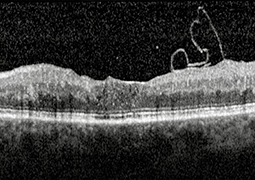
A 70-year-old woman was examined with a 10-day history of photopsia and floaters in her left eye. Her best-corrected visual acuity was 20/25 in both eyes, with a normal intraocular pressure and some nuclear sclerosis. Spectral-domain optical coherence tomography revealed a separated posterior vitreous, with a rolled internal limiting membrane flap and inner retinal dimples in the left eye. Optical coherence tomography angiography demonstrated reduced vessel density in both the superficial and deep capillary plexuses of the left fundus. Sixteen months earlier, she had received a single intravitreal Dexamethasone implant injection, due to inferotemporal branch retinal vein occlusion-related macular edema. A diagnosis of internal limiting membrane tear following an uneventful posterior vitreous detachment was reached and no treatment was recommended.

References

  1. Lattanzio R, Torres Gimeno A, Battaglia Parodi M, Bandello F. Retinal vein occlusion: current treatment. Ophthalmologica. 2011;225(3):135-143.
  2. Schmitz K, Maier M, Clemens CR, et al. Reliability and safety of intravitreal Ozurdex injections: The ZERO study. Der Ophthalmologe. 2014 Jan;111:44-52.
  3. ALBalawi HB, Alali NM, Altemani AH, et al. Anterior segment migration of intravitreal dexamethasone implant in a patient with scleral fixation intraocular lens implant: a case report. J Surg Case Rep. 2024 Mar 8;2024(3):rjae121.
  4. Ambiya V, Shankar S, Kumar A, Srujana D. Toxoplasma retinochoroiditis following intravitreal dexamethasone implant injection for diabetic macular edema: A rare complication. Ocul Immunol Inflamm. 2024 Aug;32(6):1121-1123.
  5. Rajesh B, Zarranz-Ventura J, Fung AT, et al. Safety of 6000 intravitreal dexamethasone implants. Br J Ophthalmol. 2020 Jan;104(1):39-46.
  6. Bovey EH, Uffer S. Tearing and folding of the retinal internal limiting membrane associated with macular epiretinal membrane. Retina. 2008 Mar;28(3):433-440.
  7. Durrani AF, Hyde RA, Johnson MW. Large internal limiting membrane tears: Prevalence, clinical characteristics, and surgical utility. Am J Ophthalmol. 2023 Jan;245:115-125.
  8. Yeo JH, Kim JG. Bare retinal nerve fiber layer and internal limiting membrane tear associated with epiretinal membrane: OCT findings and visual outcomes. Graefes Arch Clin Exp Ophthalmol. 2020 Jul;258(7):1379-1387.
  9. Jain M, Narayanan R, Barik B, Sahoo NK, Raval V, Reddy NG. Internal limiting membrane detachment in acute central retinal artery occlusion: Clinical features, multimodal imaging, outcomes & prognostic biomarker. Int J Retina Vitreous. 2022 Dec;8(1):86.
  10. Gelman R, Stevenson W, Prospero Ponce C, Agarwal D, Christoforidis JB. Retinal damage induced by internal limiting membrane removal. J Ophthalmol. 2015;2015:939748.
  11. Liu J, Chen Y, Wang S, Zhang X, Zhao P. Evaluating inner retinal dimples after inner limiting membrane removal using multimodal imaging of optical coherence tomography. BMC Ophthalmol. 2018 Jun 27;18(1):155.
  12. Bringmann A, Unterlauft JD, Wiedemann R, Barth T, Rehak M, Wiedemann P. Two different populations of Müller cells stabilize the structure of the fovea: an optical coherence tomography study. Int Ophthalmol. 2020 Nov;40:2931-2948.
  13. Navajas EV, Schuck N, Govetto A, et al. En face optical coherence tomography and optical coherence tomography angiography of inner retinal dimples after internal limiting membrane peeling for full-thickness macular holes. Retina. 2020 Mar;40(3):557-566.
  14. Spaide RF. “Dissociated optic nerve fiber layer appearance” after internal limiting membrane removal is inner retinal dimpling. Retina. 2012 Oct;32(9):1719-1726.
  15. Avci R, Mavi Yildiz A, Yilmaz S. Evaluation of inner retinal dimples and internal limiting membrane flap configuration after temporal inverted ILM flap technique. Eur J Ophthalmol. 2021 Mar;31(2):649-655.
  16. Nakamura T, Murata T, Hisatomi T, et al. Ultrastructure of the vitreoretinal interface following the removal of the internal limiting membrane using indocyanine green. Curr Eye Res. 2003 Dec;27(6):395-399.
  17. Eckardt C, Eckardt U, Groos S, Luciano L, Reale E. Removal of the internal limiting membrane in macular holes. Clinical and morphological findings. Der Ophthalmologe: Zeitschrift der Deutschen Ophthalmologischen Gesellschaft. 1997 Aug 1;94(8):545-551.
  18. Geck U, Pustolla N, Baraki H, Atili A, Feltgen N, Hoerauf H. Posterior vitreous detachment following intravitreal drug injection. Graefes Arch Clin Exp Ophthalmol. 2013 Jul;251(7):1691-1695.
  19. Hussnain SA, Sharma T, Hood DC, Chang S. Schisis of the retinal nerve fiber layer in epiretinal membranes. Am J Ophthalmol. 2019 Nov;207:304-312.
  20. Kishi S, Numaga T, Yoneya S, Yamazaki S. Epivascular glia and paravascular holes in normal human retina. Graefes Arch Clin Exp Ophthalmol. 1986;224(2):124-130.