
2023 - 6 Issue

Original article

Comparison of Three Methods of Tonometry in Patients With Inactive Thyroid-Associated Orbitopathy

Full Text links

Summary

Introduction: Intraocular pressure (IOP) measurement in patients with thyroid-associated orbitopathy (TAO) can be difficult and misleading, particularly in patients with diplopia and eye deviation (esotropia or hypotropia). However, when measuring IOP, it is also necessary to pay sufficient attention to TAO patients without diplopia in primary gaze direction and without motility disorder that might not be readily apparent.

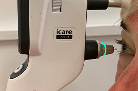
Purpose: The aim of this study was to evaluate the accuracy of measurement of intraocular pressure (IOP) using three different types of tonometers: the rebound tonometer (iCARE), the Goldmann applanation tonometer (GAT) and the non-contact airpuff tonometer (NCT) in patients with inactive TAO.

Materials and Methods: A total of 98 eyes of 49 adult patients with TAO were examined. The study group included 36 females and 13 males, with an age range of 19–70 years and a median age of 55.0. All the patients had evidence of thyroid disease, a history of mild to moderate TAO, no clinical signs or symptoms of active disease, and no diplopia in direct gaze direction. In addition to a comprehensive eye examination, all the patients underwent measurement of intraocular pressure with three tonometers: NCT, iCARE, and GAT. The measurements with these three devices were compared.

Results: The mean IOP was 18.1 ± 2.4 mmHg (range 13–25 mmHg) with GAT, 22.3 ±5.0 mmHg (range 13–35 mmHg) with NCT, and 18.0 ±2.4 mmHg (range 13.3–26 mmHg) with iCARE. The mean difference between the GAT and iCARE measurements (using the Bland-Altman analysis) was -0.1
±1.16 mmHg (limits of agreement -2.4 to 2.1). The mean difference between the GAT and NCT measurements was 4.2 ±3.6 mmHg (limits of agreement -2.8 to 11.2). The mean difference between the iCARE and NCT measurements was -4.3 ±3.7 mmHg (limits of agreement -11.6 to 2.9). No significant difference was found between GAT and iCARE (p = 1.000). However, there was a significant difference between GAT and NCT (p < 0.0001), as well as between iCARE and NCT (p < 0.0001).

Conclusions: In patients with TAO, NCT significantly overestimates IOP values compared to the GAT and ICare. By contrast, the iCARE rebound tonometer provides IOP measurements comparable to the gold standard GAT in these patients.

References

  1. Behrouzi Z, Rabei HM, Azizi F et al. Prevalence of open-angle glaucoma, glaucoma suspect, and ocular hypertension in thyroid-related immune orbitopathy. J Glaucoma. 2007;16(4):358-362.
  2. Ohtsuka K, Nakamura Y. Open-angle glaucoma associated with Graves disease. Am J Ophthalmol. 2000;129(5):613-617.
  3. Karhanova M, Kalitova J, Kovar R et al. Ocular hypertension in patients with active thyroid-associated orbitopathy: a predictor of disease severity, particularly of extraocular muscle enlargement. Arch Clin Exp Ophthalmol. 2022;260(12):3977-3984.
  4. Cockerham KP, Pal C, Jani B et al. The prevalence and implications of ocular hypertension and glaucoma in thyroid-associated orbitopathy. Ophthalmology 1997;104(6):914-917.
  5. Kuebler AG, Wiecha C, Reznicek L et al. Comparison of different devices to measure the intraocular pressure in thyroid-associated orbitopathy. Graefe's Arch Clin Exp Ophthalmol 2019;257:2025-2032.
  6. Karhanová M. Glaukom a oční hypertenze při endokrinní orbitopatii. In: Sekundární glaukomy, 1. Vydání, Mladá fronta a.s. 2016;137-157.
  7. Karhanová M, Kovář R, Fryšák Z et al.: Postižení okohybných svalů u pacientů s endokrinní orbitopatií [Extraocular Muscle Involvement in Patients with Thyroid-associated Orbitopathy]. Cesk Slov Oftalmol. 2014;70(2):66-71. Czech.
  8. Fishman DR, Benes SC. Upgaze intraocular pressure changes and strabismus in Graves' ophthalmopathy. Clin Neuroophthalmol. 1991;11(3):162-165.
  9. Herzog D, Hoffmann R, Schmidtmann I et al. Is gaze-dependent tonometry a useful tool in the differential diagnosis of Graves' ophthalmopathy? Graefes Arch Clin Exp Ophthalmol 2008;246(12):1737-1741.
  10. Nardi M, Bartolomei MP, Romani A, Barca L. Intraocular pressure changes in secondary positions of gaze in normal subjects and in restrictive ocular motility disorders. Graefes Arch Clin Exp Ophthalmol 1988;226(1):8-10.
  11. Gomi CF, Yates B, Kikkawa DO et al. Effect on intraocular pressure of extraocular muscle surgery for thyroid-associated ophthalmopathy. Am J Ophthalmol 2007;144(5):654-657.
  12. Kikkawa DO, Cruz RC Jr, Christian WK et al. Botulinum A toxin injection for restrictive myopathy of thyroid-related orbitopathy: effects on intraocular pressure. Am J Ophthalmol 2003;135(4):427-31.
  13. Karabulut GO, Kaynak P, Altan C et al. Corneal biomechanical properties in thyroid eye disease. Kaohsiung J Med Sci 2014;30(6):99- 304.
  14. Pérez-López M, Ting DSJ, Lanzagorta-Aresti A et al. Influence of corneal biomechanical properties on intraocular pressure measurement in different types of Graves’ orbitopathy. Eur J Ophthalmol. 2023;33(1):567-573.
  15. Flemmons MS, Hsiao YC, Dzau J, Asrani S, Jones S, Freedman SF. Icare rebound tonometry in children with known and suspected glaucoma. J AAPOS. 2011;15(2):153-157.
  16. Nakakura S. Icare® rebound tonometers: review of their characteristics and ease of use. Clin Ophthalmol. 2018;12:1245-1253.
  17. Pakrou N, Gray T, Mills R, Landers J, Craig J. Clinical comparison of the Icare tonometer and Goldmann applanation tonometry. J Glaucoma. 2008;17(1):43-47.
  18. Stoor K, Karvonen E, Ohtonen P, Liinamaa MJ, Saarela V. Icare versus Goldmann in a randomised middle-aged population: The influence of central corneal thickness and refractive errors. Eur J Ophthalmol. 2021;31(3):1231-1239.
  19. Hladíková E, Pluháček F, Marešová K. Porovnání měření nitroočního tlaku ICARE PRO tonometrem a Goldmanovým aplanačním tonometrem [Comparison of measurement of intraocular pressure by ICARE PRO® tonometer and Goldman applanation tonometer]. Cesk Slov Oftalmol. 2014 70(3):90-93. Czech.