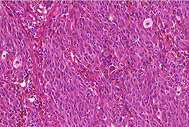
In intraocular tumors, diagnosis is usually based on clinical examination and imaging without the need for invasive surgery or tissue sampling. The diagnosis can be confirmed by biopsy, however, in the case of intraocular malignancy, the biopsy is considered controversial. Due to the development of uveal melanoma cytogenetic prognostics and the progression in generalised uveal melanoma treatment, intraocular melanoma biopsy is becoming increasingly important. Diagnostic biopsy of intraocular tumors is indicated in cases of diagnostic uncertainty for findings with conflicting non-invasive test results and for small melanocyte lesions. Tumor prognostic biopsy is performed to obtain a tissue sample for tumor cytogenetic testing, which can help to determine the prognosis and specific metastatic risk of the patient.
For anterior segment tumors, anterior chamber fluid sampling, thin-needle iris biopsy, punch biopsy, surgical biopsy or biopsy using vitrectomy may be used. For posterior segment tumors, procedures include transscleral or transretinal thin-needle biopsy, vitrectomy-assisted biopsy, punch biopsy, endoresection or transscleral exoresection. Complications of intraocular melanoma biopsy include too small or non-valuable sample collection, intra-tumoral heterogeneity, intra-ocular trauma and induction of intraocular or extraocular tumor dissemination.
- Uveal Melanoma Biopsy. A Review
- Visual Functions after Implantation of Acrysof Monofocal Intraocular Lenses
- Evaluation of Patients Presenting to the Ophthalmology Department of a Tertiary Hospital for Nonemergency Reasons During the Covid-19 Pandemic
- Changed Eye Functions and Quality of Life of Seniors with Diabetic Retinopathy
- Can Visual Function Be Affected by an Open Foramen Ovale?
- Late Functional and Morphological Findings after Methylalcohol Poisoning

