
2020 - 5 Issue

Others

Highlights of Hypertensive and Normotensive Glaucoma

Full Text links

Summary

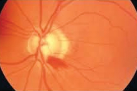
The paper presents the up-to-date overview of pathogenesis, functional and structural changes in normotensive glaucoma (NTG) and its differences from hypertensive glaucoma (HTG).

The autors point out new facts that distinguish both diagnostic groups. In the first place are the results of OCT angiography, which verify the pathology of NTG to the anterior part of optic nerve.

Our findings confirmed that vascular component (VD) is more involved in changes of visual field than in perfusion parameters, especially in arteria ophtalmica (AO). Perfusion in arteria centralis retinae (ACR) does not play a significant role in NTG changes in the visual field.

VD has very little effect on changes in visual field in HTG. Similarly, the retinal nerve fiber layer (RNFL) for changes in the visual field. Howerver, VD is moderately influenced by changes in RNFL. It should be emphasized that we compared the sum of sensitivity in the central part of the visual field (0-22 degrees) with RNFL and VD. In NTG, the anterior part of the optic nerve is altered. Mainly VD contributes to visual field changes in NTG. It is also important to note that when the intraocular pressure (IOP) increased above 20 mm Hg, the macular and papillary VD was significantly reduced.

Antiglaucomatous treatment with prostaglandins and beta-blockers is essential for the reduction of IOP in HTG. This reduction shoud be bellow 20 mm Hg, in eyes with thinner cornea the decrease in IOP should be more pronounced. It does not matter which antiglaucoma treatment was used. However, it should be noted that prostaglandins have a greater effect on disease progression, but the greater protective effect on the visual field have beta-blockers. Neuroprotectives should be recommended systemically in patients with HTG.

When treating NTG, it is important to maintain blood flow of the posterior pole of the eye, but mainly of the anterior part of the optic nerve. Prostaglandins are not suitable in NTG patients, although their effect on IOL reduction is high. Beta-blockers (betaxolol and carteol) and brimonidine are most suitable. Corneal thickness has no effect on disease progression.

References

  1. Our findings confirmed that the vascular component of VD contributes to changes in the visual fields in NTG to a greater degree than perfusion parameters, especially in the AO. Perfusion in the ACR does not play a significant role in changes in the visual field in NTG.
  2. VD has very little influence on changes in the visual fields in HTG. The same applies to the retinal nerve fibre layer (RNFL) on changes in the visual field. However, VD has a medium strength influence on changes in the RNFL. Here it is necessary to emphasise that we compared the sum of sensitivities in the central part of the visual field (0-22 degrees) with the RNFL and VD. In NTG an alteration of the anterior part of the optic nerve takes place. VD has a greater share in changes in the visual fields in NTG. However, it is important to state that when intraocular pressure (IOP) increased above 20 mmHg, macular and papillary VD was significantly reduced.
  3. Antiglaucomatous treatment by prostaglandins and beta-blockers is of fundamental significance for reducing IOP in HTG. This reduction should be below 20 mmHg, in eyes with a thinner cornea the reduction of IOP should be more pronounced. It is not of fundamental significance as to which antiglaucomatous agents are used. It is nevertheless necessary to point to the fact that prostaglandins have a greater influence on the progression of the pathology. Beta-blockers have a greater protective effect on the visual field. In patients with HTG it is appropriate to recommend neuroprotective agents systemically.
  4. In the treatment of NTG it is important to preserve blood perfusion of the posterior pole of the eye, but above all of the anterior part of the optic nerve. Prostaglandins are not suitable for patients with NTG, even if they have a large influence on reducing IOP. The most appropriate drugs are beta-blockers (betaxolol and carteol) and brimonidine. Corneal thickness has no influence on the progression of the pathology.
  5. Kuerten D, Fuest M, Bienert M. et al. Ocular hemodynamics in Acute Nonarteritic Anterior Ischemic Optic Neuropathy Compared with normal tension glaucoma. J Glaucoma. 2019;28: 334–340.
  6. Betaxolol, brimonidine and carteolol in the treatment of normotensive glaucomas Following on from the study “The Influence of Prostaglandins and Beta-blockers on the Progression of Hypertensive and Normotensive Glaucomas”, we were interested in whether any ophthalmological drugs from the range of beta-blockers had a different influence than brimonidine on the progression of NTG [12]. We included 30 patients with NTG in the cohort. The first group was composed of twenty eyes of ten patients (mean age 58.5 years) treated with betaxolol, the second group also of twenty eyes of ten patients (mean age 62.6 years) treated with brimonidine, and the third group of the same number (mean age 61.1 age) treated with carteolol. We examined the visual field with a fast threshold glaucoma program on the instrument Medmont M700. We compared PD of the visual field within the range of three years. In PD we did not record a statistically significant difference in any of the groups. We also did not determ
  7. Zaková M, Lestak J, Fus M, Maresova K. OCT Angiography and Visual Field in Hypertensive and Normotensive Glaucoma. Biomed Pap Med Fac Univ Palacky Olomouc Czech Repub. 2020;164 (in press)
  8. Ma ZW, Qiu WH, Zhou DN, Yang WH, Pan XF, Chen H. Changes in vessel density of the patients with narrow antenior chamber after an acute intraocular pressure elevation observed by OCT angiography. BMC Ophthalmol. 2019;19:132. doi: 10.1186/s12886-019- 1146-6.
  9. Cheng HC, Chan CM, Yeh SI, Yu JH, Liu DZ. The hemorheological mechanisms in normal tension glaucoma. Curr Eye Res. 2011;36:647–653.
  10. Lestak J, Jiraskova N, Zakova M, Stredova M. Normotensive glaucoma. Biomed Pap Med Fac Univ Palacky Olomouc Czech Repub. 2018;162:272–275.
  11. Lestak J, Nutterova E, Bartosova L, Rozsival P. The Visual Field in Normal Tension and Hyper Tension Glaucoma. IJSR. 2014;3:49–51.
  12. Maresova K, Lestak J, Fus M, Weisova I. Effect of prostaglandins and beta blockers on progression of hypertensive and normotensive glaucomas. Biomed Pap Med Fac Univ Palacky Olomouc Czech Repub. X:X | DOI: 10.5507/bp.2020.011
  13. Lestak J, Fus M: Neuroprotection in glaucoma – a review of electrophysiologist. Exp Ther Med. 2020;18: DOI: 10.3892/etm.2020.8509
  14. Lešták J, Rozsíval P. The Influence of Corneal Thickness on Progression of Hypertensive Glaucoma. J Clin Exp Ophthalmol. 2012;3:8:http://dx.doi.org/10.4172/2155-9570.1000245.
  15. Nutterova E, Maresova K, Lestak J. The Influence of Central Corneal Thickness on Progression of Normotensive Glaucoma. Biomed Pap Med Fac Univ Palacky Olomouc Czech Repub. 2019 Nov 18. doi: 10.5507/bp.2019.057.
  16. Lešták J, Fus M, Weissova I, Marešova K. Betaxolol, brimonidin and carteolol in the therapy normotensive glaucoma. Cesk Slov Oftalmol. 2020;76:94–97.