
2020 - 3 Issue

Original article

Changes of the Foveal Avascular Zone and Macular Microvasculature within the Framework of OCT Angiography Examination in Young Patients with Type 1 Diabetes (Pilot Study)

Full Text links

Summary

Aim: Learn about the development and changes in foveal avascular zone (FAZ) and vascularity of retina in the surrounding zone, depending on the duration in young diabetic patients type 1 (T1DM).

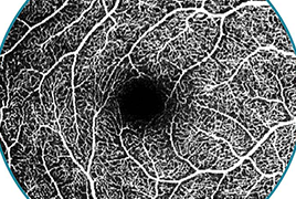
Methods: As part of regular one-year examinations of young T1DM patients at the Eye Clinic of the University Hospital Královské Vinohrady in Prague (Czech Republic, EU) from January to December 2019, OCT angiography using the device Spectralis (Heidelberg Engineering) was included. Forty patients aged 18 to 30 years were examined, median 21 years. T1DM was diagnosed in childhood and lasted for more than 10 years. At the same time, a control group of forty individuals of similar age, without metabolic and other general disease was examined, normal visual acuity and physiological fundoscopic finding were obligatory. The FAZ size was evaluated in both groups (using built-in function "Draw Region"), also its shape, density decrease and change in character of vascularity of the retina was assessed.

Results: In the control group, the FAZ area ranged from 0.06 to 0.4 mm², with an average of 0.253 ± 0.092 mm² and a median of 0.27 mm². It was not affected by a fundamental change in its round shape and the surrounding capillary netting was regular and reasonably dense. In T1DM patients, the FAZ area was in a wider range, from 0.05 to 0.64 mm², an average of 0.300 ± 0.132 mm², and a median of 0.31 mm². The difference in FAZ across-the-board evaluation was statistically significant (p = 0, 009). Diabetic preretinopathy (DpR) was defined by the irregularity of the capillary density up to the manifestation of non-perfusion, in 61% of cases the size of the FAZ was changed. In diabetic retinopathy (DR) there was always an irregularity of the FAZ shape with its enlargement, manifestation of non-perfusion, capillary dilatation and rare microaneurysms.

Conclusion: Changes in FAZ size corresponded to the stage of T1DM on the fundoscopic finding of the eye depending on its duration. The initial increased amount of foveal capillaries, which resulted in decreased FAZ area, was followed by a gradual decrease in capillaries and increased FAZ area, consistent with the manifestations of DpR. It was accompanied by a change in capillary density in macula to eventual non-perfusion. On the contrary, the increase in the FAZ area and its irregularity accompanied by non-perfusion of the capillary net and microaneurysms corresponded to the development of DR already.

References

  1. Němec P. Trendy soudobé oftalmologie. Svazek 10. Praha: Galén; 2017. OCT angiografie; s. 33–50.
  2. Krásný J, Anděl M, Brunnerová R, et al. The contrast sensitivity test in early detection of ocular changes in the relation to the type 1 diabetes mellitus compensation in children, teenegers, and young adults. Recent Pat Inflamm Alergy Drug Discov. 1; 2007; 232–236.
  3. Krásný J, Vosáhlo J, Čeledová J, et al. Preretinopatie u mladistvých s diabetem I. typu z pohledu citlivosti na kontrast a koherentní tomografie (Pilotní studie). Ces Slov Oftal. 2014;70:123–130.
  4. Kalvodová B, Sosna T, Ernest J, et al. Doporučené postupy pro diagnostiku a léčbu diabetické retinopatie. Ces Slov Oftal. 2016;72:226–233.
  5. Sosna T, Bouček T, Fišer I. Diabetická retinopatie. Praha: Jiří Cendelín; 2001. 255.
  6. Cunha-Vaz JG. Pathophysiology of diabetic retinopathy. British Journal of Ophthalmology. 1978;62(6):351–5.
  7. Cunha-Vaz JG, Goldberg MF, Vygantas C, Noth J. Early detection of retinal involvement in diabetes by vitreous fluorophotometry. Ophthalmology. 1979;86(2):264–75.
  8. Tomčíková D, Gerinec A. Osobitosti diabetické retinopatie u dětí. Ces Slov Oftal. 2001;57:266–276.
  9. Filouš A, Rychnová J, Vavřinec J. Rozšířená klasifikace diabetické retinopatie u dětí a mladistvých. Cs Pediatr. 1992;47:453–455.
  10. Verougstraete C, Toussaint D, De Schepper J, Haentjens M, Dorchy H. First microangiographic abnormalities in childhood diabetes - types of lesions. Graefe’s archive for clinical and experimental ophthalmology. 1991;229(1):24–32.
  11. Klement UM, Freyler H. Diabetische Präretinopathie. Klin Mbl Augenheilk. 1980;176:313–316. German.
  12. Enders C, Baeuerle F, Lang GE, Dreyhaupt J, Lang GK, Loidl M, et al. Comparison between Findings in Optical Coherence Tomography Angiography and in Fluorescein Angiography in Patients with Diabetic Retinopathy. Ophthalmologica. 2019 May 28;1–6.
  13. Mihailovic N, Eter N, Alnawaiseh M. Foveal avascular zone and OCT angiography. An overview of current knowledge. Ophthalmologe. 2019 Jul;116(7):610–6.
  14. Vujosevic S, Toma C, Villani E, Gatti V, Brambilla M, Muraca A, et al. Early Detection of Microvascular Changes in Patients with Diabetes Mellitus without and with Diabetic Retinopathy: Comparison between Different Swept-Source. J Diabetes Res. 2019;2019:2547216.
  15. Xiao H, Liu X, Liao L, Tan K, Ling Y, Zhong Y. Reproducibility of Foveal Avascular Zone and Superficial Macular Retinal Vasculature Measurements in Healthy Eyes Determined by Two Different Scanning Protocols of Optical Coherence Tomography Angiography. Ophthalmic Res. 2019 Oct 16;1–8.
  16. Ashraf M, Nesper PL, Jampol LM, Yu F, Fawzi AA. Statistical Model of Optical Coherence Tomography Angiography Parameters That Correlate With Severity of Diabetic Retinopathy. Invest Ophthalmol Vis Sci. 2018 Aug 1;59(10):4292–8.
  17. Ishii H, Shoji T, Yoshikawa Y, Kanno J, Ibuki H, Shinoda K. Automated Measurement of the Foveal Avascular Zone in Swept-Source Optical Coherence Tomography Angiography Images. Transl Vis Sci Technol. 2019 May;8(3):28.
  18. Hsu ST, Ngo HT, Stinnett SS, Cheung NL, House RJ, Kelly MP, et al. Assessment of Macular Microvasculature in Healthy Eyes of Infants and Children Using OCT Angiography. Ophthalmology. 2019 Jul 15;
  19. Gomez-Ulla F, Cutrin P, Santos P, Fernandez M, Abraldes M, Abalo-Lojo JM, et al. Age and gender influence on foveal avascular zone in healthy eyes. Exp Eye Res. 2019 Oct 22;189:107856.
  20. Gołębiewska J, Biała-Gosek K, Czeszyk A, Hautz W. Optical coherence tomography angiography of superficial retinal vessel density and foveal avascular zone in myopic children. PLoS ONE. 2019;14(7):e0219785.
  21. Lynch G, Romo JSA, Linderman R, Krawitz BD, Mo S, Zakik A, et al. Within-subject assessment of foveal avascular zone enlargement in different stages of diabetic retinopathy using en face OCT reflectance and OCT angiography. Biomed Opt Express. 2018 Dec 1;9(12):5982–96.
  22. Wang B, Camino A, Pi S, Guo Y, Wang J, Huang D, et al. Three-dimensional structural and angiographic evaluation of foveal ischemia in diabetic retinopathy: method and validation. Biomed Opt Express. 2019 Jul 1;10(7):3522–32.
  23. Vujosevic S, Muraca A, Alkabes M, Villani E, Cavarzeran F, Rossetti L, et al. Early microvascular and neural changes in patients with type 1 and type 2 diabetes mellitus without clinical signs of diabetic retinopathy. Retina. 2019 Mar;39(3):435–45.
  24. Conrath J, Giorgi R, Raccah D, Ridings B. Foveal avascular zone in diabetic retinopathy: quantitative vs qualitative assessment. Eye (Lond). 2005 Mar;19(3):322–6.
  25. Bates NM, Tian J, Smiddy WE, Lee W-H, Somfai GM, Feuer WJ, et al. Relationship between the morphology of the foveal avascular zone, retinal structure, and macular circulation in patients with diabetes mellitus. Sci Rep. 2018 Mar 29;8(1):5355.
  26. Lavia C, Feldman-Billard S, Erginay A, Guerin O, Virgili G, Gaudric A, et al. Rapid macular capillary loss in patients with uncontrolled type 1 diabetes. Retina. 2019 May 8;
  27. Lu Y, Simonett JM, Wang J, Zhang M, Hwang T, Hagag AM, et al. Evaluation of Automatically Quantified Foveal Avascular Zone Metrics for Diagnosis of Diabetic Retinopathy Using Optical Coherence Tomography Angiography. Invest Ophthalmol Vis Sci. 2018 May 1;59(6):2212–21.
  28. Sousa DC, Leal I, Moreira S, do Vale S, Silva-Herdade AR, Dionisio P, et al. Optical coherence tomography angiography study of the retinal vascular plexuses in type 1 diabetes without retinopathy. Eye (Lond). 2019 Jul 4;
  29. Sun Z, Tang F, Wong R, Lok J, Szeto SKH, Chan JCK, et al. OCT Angiography Metrics Predict Progression of Diabetic Retinopathy and Development of Diabetic Macular Edema: A Prospective Study. Ophthalmology. 2019 Jun 26;
  30. AttaAllah HR, Mohamed AAM, Ali MA. Macular vessels density in diabetic retinopathy: quantitative assessment using optical coherence tomography angiography. Int Ophthalmol. 2019 Aug;39(8):1845–59.
  31. Balaratnasingam C, Inoue M, Ahn S, McCann J, Dhrami-Gavazi E, Yannuzzi LA, et al. Visual Acuity Is Correlated with the Area of the Foveal Avascular Zone in Diabetic Retinopathy and Retinal Vein Occlusion. Ophthalmology. 2016 Nov;123(11): 2352–67.
  32. Moein H-R, Novais EA, Rebhun CB, Cole ED, Louzada RN, Witkin AJ, et al. Optical coherence tomography angiography to detect macular capillary ischemia in patients with inner retinal changes after resolved diabetic macular edema. Retina. 2018 Dec;38(12):2277–
  33. Kapsala Z, Pallikaris A, Mamoulakis D, Moschandreas J, Bontzos G, Tsilimbaris M. Perifoveal capillary network quantification in young diabetic patients with subclinical or no retinopathy. Can J Ophthalmol. 2018 Jun;53(3):199–206.
  34. Rosen RB, Andrade Romo JS, Krawitz BD, Mo S, Fawzi AA, Linderman RE, et al. Earliest Evidence of Preclinical Diabetic Retinopathy Revealed Using Optical Coherence Tomography Angiography Perfused Capillary Density. Am J Ophthalmol. 2019 Jul;203:103–15.
  35. Sacconi R, Casaluci M, Borrelli E, Mulinacci G, Lamanna F, Gelormini F, et al. Multimodal Imaging Assessment of Vascular and Neurodegenerative Retinal Alterations in Type 1 Diabetic Patients without Fundoscopic Signs of Diabetic Retinopathy. J Clin Med. 2019 Sep 8;8(9).
  36. Takase N, Nozaki M, Kato A, Ozeki H, Yoshida M, Ogura Y. Enlargement of foveal avascular zone in diabetic eyes evaluated by en face optical coherence tomography angiography. Retina. 2015 Nov;35(11):2377–83.
  37. de Carlo TE, Chin AT, Bonini Filho MA, Adhi M, Branchini L, Salz DA, et al. Detection of microvascular changes in eyes of patients with diabetes but not clinical diabetic retinopathy using optical coherence tomography angiography. Retina. 2015 Nov;35(11):2364–70.
  38. Gildea D. The diagnostic value of optical coherence tomography angiography in diabetic retinopathy: a systematic review. Int Ophthalmol. 2019 Oct;39(10):2413–33.
  39. Ruminski D, Sikorski BL, Bukowska D, Szkulmowski M, Krawiec K, Malukiewicz G, et al. OCT angiography by absolute intensity difference applied to normal and diseased human retinas. Biomedical optics express. 2015;6(8):2738–54.
  40. Miwa Y, Murakami T, Suzuma K, Uji A, Yoshitake S, Fujimoto M, et al. Relationship between functional and structural changes in diabetic vessels in optical coherence tomography angiography. Scientific reports. 2016;6:29064.
  41. Xu X, Chen C, Ding W, Yang P, Lu H, Xu F, et al. Automated quantification of superficial retinal capillaries and large vessels for diabetic retinopathy on optical coherence tomographic angiography. J Biophotonics. 2019 Nov;12(11):e201900103.
  42. Munk MR, Giannakaki-Zimmermann H, Berger L, Huf W, Ebneter A, Wolf S, et al. OCT-angiography: A qualitative and quantitative comparison of 4 OCT-A devices. PloS one. 2017;12(5): e0177059.
  43. Inanc M, Tekin K, Kiziltoprak H, Ozalkak S, Doguizi S, Aycan Z. Changes in Retinal Microcirculation Precede the Clinical Onset of Diabetic Retinopathy in Children with Type 1 Diabetes Mellitus. Am J Ophthalmol. 2019 Apr 19;
  44. Simonett JM, Scarinci F, Picconi F, Giorno P, De Geronimo D, Di Renzo A, et al. Early microvascular retinal changes in optical coherence tomography angiography in patients with type 1 diabetes mellitus. Acta Ophthalmol. 2017 Dec;95(8):751–5.
  45. Niestrata-Ortiz M, Fichna P, Stankiewicz W, Stopa M. Enlargement of the foveal avascular zone detected by optical coherence tomography angiography in diabetic children without diabetic retinopathy. Graefes Arch Clin Exp Ophthalmol. 2019 Apr;257(4): 689–97.