
2020 - 1 Issue

Original article

The Importance of Pet/Ct Examination in Patients With Malignant Uveal Melanoma

Full Text links

Summary

Introduction: Diagnostic and therapeutic management of the patient with malignant uveal melanoma (MMU) is subject to ongoing efforts to innovate. PET/CT (Positron Emission Tomography / Computed Tomography) examination is important in both diagnosis and metastases.

Material and methods: Evaluation of the importance of PET/CT examination in the group of patients diagnosed with MMU in the period 12.1.2016 to 6.12.2018. All patients with a diagnosis of secondary retinal detachment, suspected uveal melanoma, underwent standard examinations to detect possible metastases (liver ultrasound, chest X-ray). Patients for whom a stereotactic radiosurgery solution was planned due to the stage of the disease this examination was to exclude metastasis in the liver or lungs. PET/CT examination is part of the protocol within the exclusion criteria for treatment with stereotactic radiosurgery in one day session surgery.

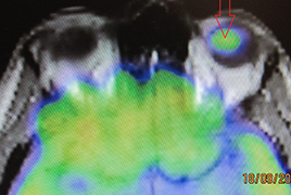
Results: In the group of 84 patients, 47 women (56 %) and 37 men (44 %) were aged between 26 and 90 years. Their average age was 61.4 years. The median group was 64 years, modus 65 years. Of 84 patients, 79 (94 % of cases) had a diagnosis of C69.3 (choroidal melanoma) and 5 patients (6 % of cases) had a diagnosis of C69.4 (ciliary body melanoma). Subsequent PET/CT examination in many patients did not reveal hypermetabolic manifestations that could involve various pathological processes, in others the radiopharmaceutical was captured in the primary tumor area of the uveal tract. Hypermetabolism in eye globe was only found in melanomas with a volume of more than 0.5 cm3. PET/CT examinations were 85, with one patient undergoing examination twice. However, in 25 patients (26 examinations), the radiopharmaceutical was taken up in places that subsequently required closer attention. The initial aim of the examination was to locate possible metastases of MMU. In the others, 3 incidents have been reported: increased metabolism in the lung and liver, thyroid and mediastinal lymph nodes. Of the 85 examinations, 26 (30.6 %) resulted in a hypermetabolic manifestation of accumulation, which was not located in the eye tract, resp. right in the eye. Two malignancies (prostatic carcinoma and rectosigmal carcinoma) have occurred in two patients. Very important was the discovery of MMU metastasis in the liver, which confirmed the important role of PET/CT examination in the management of MMU patients. The metastasis was discovered after repeated PET/CT examination.

Conclusion: PET/CT examination is a technically demanding examination and is one of the possibilities of imaging intraocular melanoma in tumors with volume more than 0.5 cm3. It is important in determining the grading and staging of the disease before radiosurgical treatment and also in detecting possible metastases after MMU treatment in cases where ultrasound or MRI examinations do not give a definite result. However, our study confirmed the significance of this examination for randomly detected 2 duplex malignancies (2.4%) and 3 incidentalomas (3.6%) in patients whose ophthalmologist diagnosed uveal melanoma and sent patients for full-body PET/CT examination.

References

  1. Almuhaideb, A., Papathanasiou, N., Bomanji, J.: 18F-FDG PET/ CT imaging in oncology. Ann Saudi Med, 31 (1); 2011: 3–13.
  2. Balasubramanya, R., Selvarajan, SK., Cox, M. et al.: Imaging of ocular melanoma metastasis. Br J Radiol, 89 (1065); 2016: 20160092.
  3. Balogova, S., Huchet, V., Kerrou, K. et al.: Detection of bronchio - loalveolar cancer by means of PET/CT and 18F-fluorocholine, and comparison with 18F-fluorodeoxyglucose. Nucl Med Commun, 31 (5); 2010: 389–97.
  4. Balogova, S., Talbot, J-N., Nataf, V. et al.: 18F-fluorodihydroxyphenylalanine vs other radiopharmaceuticals for imaging neuroendocrine tumours according to their type. Eur J Nucl Med Mol Imaging, 40 (6); 2013: 943–66.
  5. Balogova, S., Zakoun, JB., Michaud, L. et al.: Whole-body 18Ffluorocholine (FCH) PET/CT and MRI of the spine for monitoring patients with castration-resistant prostate cancer metastatic to bone: a pilot study. Clin Nucl Med, 39 (11); 2014: 951–9.
  6. Bozkurt, MF., Virgolini, I., Balogova, S. et al.: Erratum to: Guide - line for PET/CT imaging of neuroendocrine neoplasms with 68Ga- DOTA-conjugated somatostatin receptor targeting peptides and 18F-DOPA. Eur J Nucl Med Mol Imaging, 44 (12); 2017: 2150–1.
  7. Bozkurt, MF., Virgolini, I., Balogova, S. et al.: Guideline for PET/ CT imaging of neuroendocrine neoplasms with 68Ga-DOTA-conjugated somatostatin receptor targeting peptides and 18F-DOPA. Eur J Nucl Med Mol Imaging, 44 (9); 2017: 1588–601.
  8. Brovkina, AF: [Local treatment of choroidal melanoma: possibilities and limitations]. Vestn Oftalmol, 134 (4); 2018: 52–60.
  9. Cassou-Mounat, T., Balogova, S., Nataf, V. et al.: 18F-fluorocho - line versus 18F-fluorodeoxyglucose for PET/CT imaging in patients with suspected relapsing or progressive multiple myeloma: a pilot study. Eur J Nucl Med Mol Imaging, 43 (11); 2016: 1995–2004.
  10. Cohen, VML., Pavlidou, E., DaCosta, J. et al.: Staging Uveal Melanoma with Whole-Body Positron-Emission Tomography/Computed Tomography and Abdominal Ultrasound: Low Incidence of Metastatic Disease, High Incidence of Second Primary Cancers. Middle East Afr J Ophthalmol, 25 (2); 2018: 91–5.
  11. Fartoux, L., Balogova, S., Nataf, V. et al.: A pilot comparison of 18F-fluorodeoxyglucose and 18F-fluorocholine PET/CT to predict early recurrence of unifocal hepatocellular carcinoma after surgical resection. Nucl Med Commun, 33 (7); 2012: 757–65.
  12. Finger, PT., Kurli, M., Wesley, P. et al.: Whole body PET/CT imaging for detection of metastatic choroidal melanoma. Br J Ophthalmol, 88 (8); 2004: 1095–7.
  13. Furdová, A.: Nové trendy v liečbe malígneho melanómu uvey. In: Trendy soudobé oftalmologie. Galén; 2007, s. 15–35.
  14. Furdova, A., Horkovicova, K., Justusova, P. et al.: Is it sufficient to repeat LINEAR accelerator stereotactic radiosurgery in choroidal melanoma? Bratisl Lekárske Listy, 117 (8); 2016: 456–62.
  15. Furdová, A., Oláh, Z.: Malígny melanóm v uveálnom trakte. Bratislava: Asklepios; 2002, 175 s.
  16. Furdová, A., Oláh, Z.: Nádory oka a okolitých štruktúr. Brno: Akademické nakladatelství CERM; 2010, 152 s.
  17. Furdova, A., Slezak, P., Chorvath, M. et al.: No differences in outcome between radical surgical treatment (enucleation) and stereotactic radiosurgery in patients with posterior uveal melanoma. Neoplasma, 57 (4); 2010: 377–81.
  18. Furdova, A., Sramka, M.: Uveal malignant melanoma and stereo - tactic radiosurgery: Intraocular uveal melanoma and one-day session stereotactic radiosurgery at linear accelerator. Saarbrücken: LAP LAMBERT Academic Publishing; 2014, 188 p.
  19. Furdová, A., Šramka, M., Waczulíková, I. et al.: [Stereotactic Rediosurgery for Uveal Melanoma; Postradiation Complications]. Cesk Slov Oftalmol, 71 (3); 2015: 134–42.
  20. Furdova, A., Strmen, P., Waczulikova, I. et al.: One-day session LINAC–based stereotactic radiosurgery of posterior uveal melanoma. Eur J Ophthalmol, 22 (2); 2012: 226–35.
  21. Honavar, SG.: Is Collaborative Ocular Melanoma Study (COMS) still relevant? Indian J Ophthalmol, 66 (10); 2018: 1385–7.
  22. Horkovičová, K., Markus, J., Krčová, I. et al.: [The BRAF mutation and the possibilities of uveal melanoma metastasing prognostic markers identification]. Cesk Slov Oftalmol, 72 (4); 2016: 149–56.
  23. Hui, K-H., Pfeiffer, ML., Esmaeli, B.: Value of positron emission tomography/computed tomography in diagnosis and staging of primary ocular and orbital tumors. Saudi J Ophthalmol Off J Saudi Ophthalmol Soc, 26 (4); 2012: 365–71.
  24. Jorgov, L., Montravers, F., Balogova, S. et al.: Paediatric and adolescent Hodgkin lymphoma: information derived from diffuse organ uptake of 18 F-fluorodeoxyglucose on pre-treatment and on interim PET/CT. Eur J Nucl Med Mol Imaging, 43 (7); 2016: 1220–30.
  25. Murphy, G., Hussey, D., Metser, U.: Non-cutaneous melanoma: is there a role for (18)F-FDG PET-CT? Br J Radiol, 87 (1040); 2014: 20140324.
  26. Périé, S., Hugentobler, A., Susini, B. et al.: Impact of FDG-PET to detect recurrence of head and neck squamous cell carcinoma. Otolaryngol--Head Neck Surg Off J Am Acad Otolaryngol-Head Neck Surg, 137 (4); 2007: 647–53.
  27. Reddy, S., Kurli, M., Tena, LB. et al.: PET/CT imaging: detection of choroidal melanoma. Br J Ophthalmol, 89 (10); 2005: 1265–9.
  28. Seidl, Z., Burgetová, A., Hoffmannová, E. et al.: Radiologie pro studium i praxi. Praha: Grada Publishing, a.s.; 2012, 372 s.
  29. Smolkova, B., Horvathova Kajabova, V., Zmetakova, I. et al.: Role of epigenetic deregulation in hematogenous dissemination of malignant uveal melanoma. Neoplasma, 65 (6); 2018: 840–54.
  30. Strobel, K., Bode, B., Dummer, R. et al.: Limited value of 18F-FDG PET/CT and S-100B tumour marker in the detection of liver metastases from uveal melanoma compared to liver metastases from cutaneous melanoma. Eur J Nucl Med Mol Imaging, 36 (11); 2009: 1774–82.
  31. Surasi, DS., Bhambhvani, P., Baldwin, JA. et al.: 18 F-FDG PET and PET/CT patient preparation: a review of the literature. J Nucl Med Technol, 42 (1); 2014: 5–13.
  32. Talbot, J-N., Fartoux, L., Balogova, S. et al.: Detection of hepatoce - llular carcinoma with PET/CT: a prospective comparison of 18F-fluorocholine and 18F-FDG in patients with cirrhosis or chronic liver disease. J Nucl Med Off Publ Soc Nucl Med, 51 (11); 2010: 1699–706.
  33. Taus, Á., Aguiló, R., Curull, V. et al.: Impact of 18F-FDG PET/CT in the treatment of patients with non-small cell lung cancer. Arch Bronconeumol, 50 (3); 2014: 99–104.
  34. Weis, E., Salopek, TG., McKinnon, JG. et al.: Management of uve - al melanoma: a consensus-based provincial clinical practice guideline. Curr Oncol Tor Ont, 23 (1); 2016: e57-64.