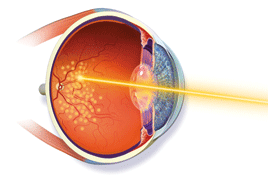
Purpose: To evaluate anatomical, functional and clinical efficacy of the pattern scanning laser (PASCAL®) photocoagulation in patients with diabetic retinopathy.
Methods: The cohort included 91 eyes in 50 patients (average age 63 years, 24 men and 26 women) treated at the Clinic of Ophthalmology, Faculty Hospital Ostrava, from 2008 to 2013. All eyes showed the signs of diabetic retinopathy (20.9% of eyes – very severe non-proliferative diabetic retinopathy (NPDR), 79.1% of eyes - proliferative diabetic retinopathy (PDR)). All the eyes were „treatment naive“. The average duration of diabetes was 18 years, average baseline HbA1c value was 8.2 %. Panretinal photocoagulation was performed with by the means of PASCAL photocoagulator – with patterns and short duration pulses. Best corrected visual acuity (BCVA), central retinal thickness (CRT), fundus photography, biomicroscopy and complications were evaluated during the minimum of 12 months follow-up period. Statistical analysis using Shapiro-Wilk a Friedman tests with p value less than 0.05 was done.
Results: Mean baseline BCVA was 0.13 logMAR. Values 0.12, 0.15 and 0.19 logMAR were observed in the follow-up intervals at the 4th, 6th and 12th month. At the 6th month visit all 91 eyes (100 %) were stabilized, and at the 12th month visit in 5 eyes (5.5 %) progression of the disease was observed. No complications were observed during the first 12 months follow up.
Conclusion: Laser photocoagulation of the retina with the use of short pulse durations and patterns in patients with DR is an effective and safe method of treatment.

