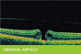
Aim: To provide a survey of the current clinical use of intraoperative optical coherence tomography (iOCT), especially in the context of our own vitreo-retinal experience.
Methods: Retrospective evaluation of casuistic cases of typical diseases of the retina, which were treated by standard pars plana vitrectomy (PPV) using iOCT integrated into an operating microscope OPMI Lumera 700 / Rescan 700 (Zeiss). Auxiliary techniques: best corrected visual acuity (BCVA) was tested on ETDRS charts, biomicroscopy was performed using a 78D lens, and optical coherence tomography (OCT) using Zeiss Cirrus equipment. Operations were carried out under retrobulbar anaesthesia by three-port 23G PPV and using a Constellation surgical unit (ALCON).
Results: Three cases of 2 females (vitreoretinal interface disorders) and 1 male (proliferating diabetic retinopathy) of average age of 63 years are reported. In the former 2 cases, the follow-up period was 3 months, and the male with diabetic retinopathy was followed for 15 months. All surgical interventions using iOCT took place without either perioperative or postoperative complications. In all cases, full anatomical success was achieved. In the former two cases, there was a marked improvement of BCVA, and in the latter case, long-term stabilization of a very good initial BCVA.
Conclusion: Surgical-microscope integrated OCT is giving the surgeon simultaneous immediate control of both surgical manipulations and OCT visualization. The results are a perfect view and information for the surgeon, and improved outcomes for the operated eye.

