
2014 - 4 Issue

Case report

Rare Case of Pathological Biomineralization of Eye Tissue

Full Text links

Summary

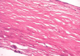
The authors have described the case of pathological biomineralization of ocular posterior chamber. Histological examination of affected eye shells revealed inflammation, oedema, dyscirculatory violations, and signs of dystrophic calcification. Structural-phase and chemical analyses of calcification have revealed that the biomineral consists of hydroxyapatite with relatively small crystallite size and defective crystal lattice. In the described case the formation of pathological biomineral in the vision organ was initiated by penetrating wound. Eye injury might have caused a hemophthalmus and chronic inflammatory reaction in the shells, these processes eventually led to the organ subatrophy and to the development of dystrophic and necrobiotic changes in the tissues. Pathological biomineralization in the affected organ developed as a type of dystrophic calcification.

References

  1. Daicker B.: Tophus-like, conglomerated, crystalline calcification of the sclera. Ophthalmologica, 1996; 210(4): 223–8.
  2. Chen KH, Li MJ, Cheng WT. et al.: Identification of monoclinic calcium pyrophosphate dihydrate and hydroxyapatite in human sclera using Raman microspectroscopy. Int J Exp Pathol, 2009; Feb;90(1):74–8.
  3. Pandey N., Guruprasad A. Choroidal osteoma with choroidal neovascular membrane successful treatment with intravitreal bevacizumab. Clinical Ophtalmology, 2010; 4, :1081–84.
  4. Bourcier T, Blain P, Massin P. et al.: Sclerochoroidal calcification associated with Gitelman syndrome. Am J Ophthalmol, 1999; Dec;128(6): 767–8.
  5. Vemuganti G.K, Honavar S.G, Jalali S.: Intraocular osseous metaplasia. A clinico-pathological study. Indian J Ophthalmol, 2002 Sep; 50(3): 183–8.
  6. Karakeshinian . [Dacrolithiasis]. Noviy armianskiy meditsinskiy jurnal [New Armenian Medical Journal]. 2011; 5 (3):71–72. (In Russ).
  7. Klug H.P., Alexander L.E. X-Ray diffraction procedures for polycrystallite and amorphous materials.-New York: JohnWiley and Sons, 1974.-965p.
  8. Moskalenko R., Romanyuk A., Danilchenko S. et al.: Morphogenetic aspects of biomineralization on the background of benign prostatic hyperplasia. Georgian medical news, 2013; 214 (1): 54–61.
  9. Danilchenko S.N., Kuznetsov V.N., Stanislavov A.S. et al.: The mineral component of human cardiovascular deposits: morphological, structural and crystal-chemical characterization. Crystal research and technology, 2013, 48 (3): 153–62.
  10. Munteanu M, Munteanu G, Giuri S, Zolog I, Motoc A.G.: Ossification of the choroid: three clinical cases and literature review of the pathogenesis of intraocular ossification. Rom J Morphol Embryol, 2013;54(3 Suppl): 871–7.
  11. Rohrbach J.M, Liesenhoff E, Steuhl K.P.: [Principles of intraocular ossification exemplified by secondary choroid ossification]. Klin Monbl Augenheilkd, 1990 Nov;197(5): 397–403.