
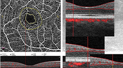

Aim: This study aims to assess and compare by means of optical coherence tomography angiography (OCTA) early modifications in the retinal layer of new-onset type 2 diabetic patients without clinical signs of diabetic retinopathy (NDR).
Materials and Methods: A total of 51 NDR patients and 53 normal individuals’ retinal capillary foveal density, superficial and deep capillary plexus, foveal avascular zone area (FAZ) parameters were evaluated with OCTA. Inner retinal layer (IRL) thickness and outer nuclear layer thickness (ONL) at the temporal and nasal margin of FAZ were measured in 26 NDR and 26 healthy subjects, using ImageJ 1.46r software.
Results: Vascular density in the superficial and deep whole image (SWIVD and DWIVD) was significantly lower in patients than in controls for right eyes (RE) and left eyes (LE), (RE SWIVD: p < 0.001, LE SWIVD: p = 0.004, RE DWIVD: p < 0.001, LE DWIVD: p = 0.028). There was no significant difference in the values of the FAZ area in the right and left eye between patient and control groups (RE: p = 0,652; LE: p = 0.509). No significant difference was found in the T/N IRL thickness between study and control groups (p = 0.552, p = 0.094, respectively). The AUC analysis for vascular densities ranged from 0.625 to 0.751.
Conclusion: Our study found that the density of blood vessels decreases before clinical signs of retinopathy appear in newly diagnosed type 2 diabetic patients.