Aim: The aim of the study was to determine the correlation between intraocular pressure (IOP) and thickness of the retinal nerve fiber layer (RNFL), and vascular density (VD) in the optic nerve.
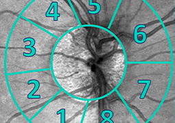
Material and methodology: IOP was greater than 21 mmHg (21–36 mmHg) in all eyes and was measured as the result of an average of three measurements with the instrument Ocular Response Analyzer (ORA, Reichert). RNFL and VD thickness (in the papillary region of 4.5 x 4.5 mm) was measured with the instrument Avanti RTVue XR (Optovue). In the case of the VD, the scan area was further separated into individual anatomical segments. In the case of corrected RNFL (RNFLc), the VD value was subtracted from the total RNFL value. The relationship of IOP to VD, RNFL and RNFLc in each peripapillary segment was determined using a Pearson’s correlation coefficient.
Results: The most significant correlation with IOP was observed for small vessel VD in a full scan (r = -0.48) and VD in the IT segment (r = -0.48). A similar correlation was observed for IOP and RNFL (r = -0.42). No statistically significant correlation was observed for RNFLc.
Conclusion: We demonstrated that VD values, specifically WI-VDs and peripapillary VDs in the IT segment, are significant markers for the early diagnosis of glaucoma.