
2017 - 1 Issue

Original article

Optical Characteristics of Myopic Corrections by Orthokeratological Contact Lenses

Full Text links

Summary

Introduction: Orthokeratology lenses present a special type of hard contact lenses, which is used especially for myopic correction. The lenses are applied during the night, when thanks to their special shape cause temporally (reversible) change of the cornea shape. During the day, there is not necessary to wear any other refractive correction. Due to the wearing regimen, there is, comparing to the classical contact lenses, higher risk of health complications. For their safe use, it is necessary to pay attention to the application of lenses of adequate parameters, follow up the care and hygiene precautions, regular controls and early treatment of possible problems. The application of contact lenses should be done by experienced and well-educated expert in contact lens fitting. In the Czech Republic, this method is practically not used. 
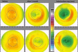
Aim: The aim of the case report was to follow up the course of concrete application of orthokeratology lenses for myopic correction from the optic (refractive) point of view, and to evaluate the optic changes in the system of the eye. In the followed up subject, there were measured subjective changes in refraction, visual acuity, corneal curvature, corneal and whole eye aberrations, and corneal thickness during the period of one month after the beginning of lens application (initial phase). The same parameters were followed-up during one-month period since the termination of lens wearing for three months (second phase). The subject was a woman, 24 years old, myopic, with the refractive error in the right eye –3.0 spherical diopters and astigmatism of –0.25 cylinder in the axis 90°, and in the left eye –2.75 spherical diopters without astigmatism; without ophthalmologic or systemic diseases. The full correction of the refractive error after the orthokeratology lenses application occurred approximately after four nights (wearing the lenses overnight). Concurrently, during the day, slight recurrence to original values was evident. With the decreased refractive error, the uncorrected visual acuity improved accordingly. Further, the flattening of the central cornea was observed, e.g. increasing of the central cornea curvature diameter (maximum change was 0.22 mm in the right and 0.28 mm in the left eye) and slight thinning. The adverse accompanying optic phenomenon was increasing of the aberrations of higher orders, which presented subjectively by decreased visual acuity under mesopic conditions. One month after the termination of contact lenses wearing, all the followed-up parameters returned to original values.

References

  1. Alharbi, A., Swarbrick, H.: The effects of overnight orthokeratology lens wear on corneal thickness. Invest Ophthalmol, Vis Sci, 44; 2003: 2518–2523.
  2. Barr, J. T., Rah, M. J., Meyers, W. et al.: Recovery of refractive error after corneal refractive therapy. Eye & Contact Lens, 30; 2004: 247–251.
  3. Gifford, P., Li, M., Lu, H. et al: Corneal nuation of overnight orthokeratology. Brit J Ophthalmol, 93; 2009: 203–208.
  4. Hiraoka, T., Okamoto, C., Ishi, Y. et al.: Recovery of corneal irregular astigmatism, ocular higher-order aberrations, and contrast sensitivity after disconti-
  5. Hiraoka, T., Okamoto, C., Ishii, Y. et al: Mesopic contrast sensitivity and ocular higher-order aberrations after overni- ght orthokeratology. Am J Ophthalmol, 145; 2008: 645–655.
  6. Hiraoka, T., Okamoto, F ., Kaji, Y. et al.: Optical quality of the cornea after sponse to mechanical stress induced by orthokeratology contact lenses. Cornea, 27; 2008: 133–141.
  7. Chan, B., Cho, P ., Cheung, S. W .: Orthokeratology practice in children in a university clinic in Hong Kong. Clinical & Experimental Optometry, 91; 2008: 453–460.
  8. Chen, D., Lam, A. K. C., Cho, P.: Posterior corneal curvature change and recovery after 6 months of overnight orthokeratology treatment. Ophthalmic & Physiological Optics: The Journal of the British College of Ophthalmic Opticians (Optometrists), 30; 2010: 274–280.
  9. Cho, P ., Collins, M., Sawano, T.: Orthokeratology Practice, A Basic Guide for Proctitioners, European Academy of Orthokeratology, 2012, 132 s.
  10. Choo, J., Caroline, P., Harlin, D.: How does the cornea change under corneal reshaping contact lenses? Eye & Contact Lens, 30; 2004: 211–213.
  11. Kobayashi, Y., Yanai, R., Chikamoto, T. et al.: Reversibility of effects of orthokeratology on visual acuity, refractive error, corneal topography, and contrast sensitivity. Eye & Contact Lens, 34; 2008: 224–228.
  12. Li, S., Kang, M., W u, S. et al.: Efficacy, Safety and Acceptability of Orthokeratology on Slowing Axial Elongation in Myopic Children by Meta-Analysis. Current Eye Research; 2015: 1–9.
  13. Lian, Y., Shen, M., Jiang, J. et al.: Vertical and horizontal thickness profiles of the corneal epithelium and Bowman’s layer after orthokeratology. Invest Ophthalmol Vis Sci, 54; 2013: 691–696.
  14. Liu YM, Xie P.: The Safety of Orthokeratology - A Systematic Review. Eye & Contact Lens, 42; 2016: 35-42.
  15. Lu, F., Simpson, T., Sorbara, L. et al.: Malleability of the ocular surface in re-
  16. Mountford, J., Ruston, D., Dave, T.: Orthokeratology: Principles and Practice, Butterworth-Heinemann, 2004, 316 s.
  17. Mountford, J.: Retention and regression of orthokeratology with time. International Contact Lens Clinic, 25; 1998: 59–64.
  18. Nichols, J. J., Marisch, M. M., Nguyen, M. et al: Overnight orthokeratology. Optometry and Vision Science: Official Publication of The American Academy of Optometry, 77; 2000: 252–259.
  19. Owens, H., Garner, L. F., Craig, J. P. et al.: Posterior Corneal Changes with Orthokeratology. Optometry & Vision Science, 81; 2004: 421–426.
  20. Potapova, N., W ang, G., Haji, S. et al: Corneal topography in corneal refractive therapy (CRT): a 1-month follow-up. Eye & Contact Lens, 30; 2004: 166–168.
  21. Reinstein, D. Z., Gobbe, M., Archer, T. J. et al.: Epithelial, stromal, and corneal pachymetry changes during orthokeratology. Optometry and Vision Science: Official Publication of the American Academy of Optometry, 86; 2009: 1006–1014.
  22. Si, J., Tang, K., Bi, H. et al.: Orthokeratology for Myopia Control: A Meta-analysis. Optometry and Vision Science: Official Publication of the American Academy of Optometry, 92; 2015: 252– 257.
  23. Sorbara, L., F onn, D., Simpson, T. et al.: Reduction of myopia from corneal refractive therapy. Optometry and Vision Science: Official Publication of the American Academy of Optometry, 82; 2005: 512–518. overnight orthokeratology. Cornea, 25; 2006: 59–63.
  24. Sridharan, R., Swarbrick, H. A.: Corneal response to short-term orthokeratology lens wear. Optometry and Vision Science: Official Publication of the American Academy of Optometry, 80; 2003: 200– 206.
  25. Stillitano, I. G., Chalita, M. R., Schor, P. et al.: Corneal changes and wavefront analysis after orthokeratology fitting test. American Journal of Ophthalmology, 144; 2007: 378–386.
  26. Sun, Y., Xu, F., Zhang, T. et al.: Orthokeratology to control myopia progression: a meta-analysis. PloS One, 10; 2015.
  27. Swabrick, H. A.: Orthokeratology review and update. Clinical & Experimental Optometry, 89; 2006: 124–143.
  28. Swarbrick, H. A., W ong, G., O’Leary, D. J.: Corneal Response to Orthokeratology. Optometry & Vision Science, 75; 1998: 791–799.
  29. Thibos, L. N., Applegate, R. A., Schwiegerling, J. T. et al: Standards for Reporting the Optical Aberrations of Eyes. J Refract Surg, 18; 2002: 265-660.
  30. Thibos L. N., Himebaugh N. L., Coe C. D.: Wavefront Refraction. In: Benjamin JW, ed.: Borish’s Clinical Refraction. 2nd Edition. Butterworth-Heinemann, 2006, s. 765-789.
  31. Wang, J., Fonn, D., Simpson, T. L. et al.: Topographical thickness of the epithelium and total cornea after overnight wear of reverse-geometry rigid contact lenses for myopia reduction. Investigative Ophthalmology & Visual Science, 44; 2003: 4742–4746.
  32. Yoon, J. H., Swarbrick, H. A.: Posterior corneal shape changes in myopic overnight orthokeratology. Optometry and Vision Science: Official Publication of the American Academy of Optometry, 90; 2013: 196–204.